Pediatric imaging has become an area of interest for researchers and clinicians working to better understand and address autism spectrum disorder (ASD). Autism is a neurodevelopmental condition characterized by challenges in social communication, restricted behaviors, and sensory sensitivities. While ASD is primarily diagnosed through behavioral assessments, imaging techniques are being explored to provide insights into brain structure and function in affected children. Here is some information about these imaging modalities and their potential contributions to studies and assessments related to autism in children.
Using Pediatric MRI for Autism Diagnosis
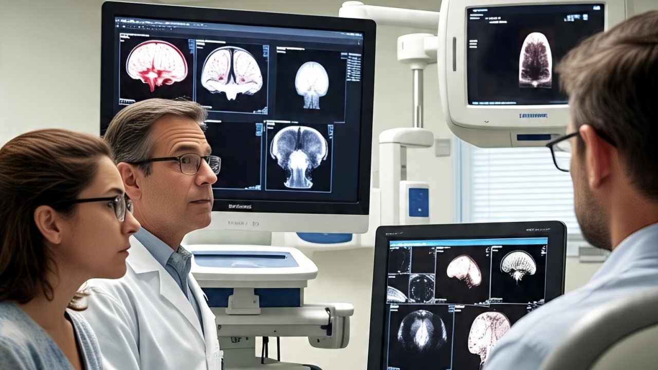
Magnetic resonance imaging (MRI) uses magnetic fields and radio waves to create detailed images of internal structures. For pediatric imaging, MRI offers a key advantage: it provides high-resolution brain images without using radiation. This makes it an excellent tool for studying brain development in children.
MRI helps researchers explore brain regions and pathways linked to functions affected by autism, such as social interaction, communication, and sensory processing. It can also identify structural differences and developmental patterns in the brain’s gray and white matter.
Preparing children for an MRI often involves extra steps. The enclosed space and long scan times can overwhelm young patients. To ease the experience, many facilities create child-friendly environments or provide sedation when necessary. Rapid imaging techniques also help shorten scan times, making the process more comfortable for kids.
Using Pediatric CT Scans to Identify Neurological Patterns
CT scans use X-rays and computer technology to create detailed cross-sectional images. While less common in autism research compared to MRIs, CT scans can help identify structural issues or injuries that may be linked to neurological symptoms. They can help rule out other conditions with similar symptoms, such as trauma or congenital abnormalities.
One concern with pediatric CT scans is radiation exposure. To address this, clinicians take steps to minimize radiation while still gathering the required information. Advances in technology have also reduced radiation levels, making CT scans safer for children.
Using Pediatric X-Rays for Health Assessments
X-ray technology, while highly accessible and commonly used, primarily serves a supporting role in the broader context of autism care. Traditional X-rays are mainly used to assess physical conditions, such as bone or chest health, rather than directly exploring brain structures.
Children with autism may need X-rays to address medical concerns, such as diagnosing or planning treatment for co-occurring conditions like gastrointestinal or musculoskeletal issues. While X-rays use ionizing radiation, healthcare professionals carefully determine their necessity. Compared to CT scans, standard X-rays use lower radiation doses, making them a quick and simple option for non-brain-related assessments.
Exploring Future Directions in Pediatric Imaging for Autism Research
Researchers are actively exploring pediatric imaging in autism studies. They use techniques like MRI to study brain differences in children, improving our understanding of neurodevelopmental patterns. These technologies advance autism insights and support clinical care. Imaging not only helps identify structural and functional brain changes but also opens doors to personalized treatment approaches. As imaging tools and research evolve, scientists continue to discover new ways to help children with autism.