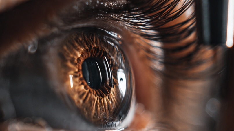
A retina specialist is an ophthalmologist with advanced training in diseases of the retina, and this doctor focuses on the back of the eye. The retina captures light. Since retinal tissue supports vision, small changes in this area may affect sight in specific ways. Here is more information on the role of a retina specialist:
Specializing in the Retina
After medical school and ophthalmology training, some doctors complete additional fellowship training in retinal care. They study complex eye disease, and they learn medical and surgical treatment methods. When the retina develops tears, swelling, or bleeding, a retina specialist evaluates the problem in detail.
These specialists manage conditions that involve fine eye structures, and their work often requires close follow-up. They treat adults and children. Since retinal disease may progress without pain, patients are typically referred after routine eye exams. A retina specialist also works with other clinicians, and this helps connect eye findings with broader health issues.
Using Extreme Precision
Retinal care involves detailed movements under magnification, and treatment decisions typically depend on subtle findings. Precision matters. Since the retina contains delicate nerve tissue, even small errors may alter how a doctor approaches care.
Some procedures take place in the office, and others are done in a surgical setting. A specialist may use:
- Laser treatment
- Eye injections
- Microsurgery
When surgery is needed, small instruments are used through tiny openings, and each step follows a planned sequence. The field is narrow. Since retinal tissue reacts to traction, fluid, and pressure, surgeons track each change closely during treatment.
Addressing Retinal Conditions
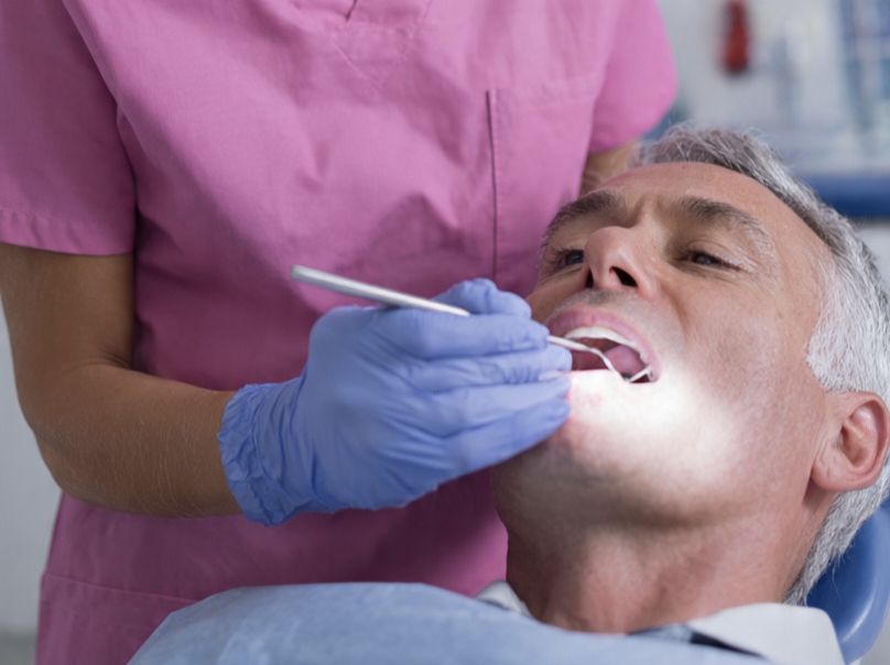
Many patients see a specialist for age-related macular degeneration, diabetic retinopathy, or retinal vein occlusion. Floaters and flashes also prompt referral. When the retina tears or detaches, prompt evaluation is typically advised.
A specialist may also treat macular holes, epiretinal membranes, and inherited retinal disorders, and each condition affects vision differently. Some diseases distort central vision. As symptoms vary by location and cause, diagnosis depends on both exam findings and imaging results.
Inflammation, trauma, and tumors also affect the retina, and these cases may require coordination with other medical teams. When unusual changes appear in the back of the eye, referral patterns can become more urgent. Early detection and timely intervention are helpful in preserving vision and preventing further complications associated with retinal conditions.
Using Diagnostic Tools
Retina specialists use imaging to study retinal layers, and these tests help measure structure and change over time. Standard tools include:
- Optical coherence tomography
- Fluorescein angiography
- Fundus photography
Some tests show swelling or leakage, and others map blood flow or document surface detail. The images are reviewed carefully by a medical professional. Since treatment plans depend on location and severity, testing guides both diagnosis and follow-up timing.
Visit a Retina Specialist
If your doctor notes retinal changes, a visit to a retina specialist may clarify the next steps, and the exam typically includes imaging. These visits are typically detailed and involve many steps. When symptoms such as new floaters, flashes, or blurred central vision appear, schedule an eye evaluation promptly.